An ACL injury occurs when there is damage to the anterior cruciate ligament, a major knee located inside the knee. The ACL is the main stabilizing ligament of the knee for sudden stopping, starting, cutting and pivoting movements. Injuries may range from disruption of a few fibers to a complete tear of the ligament. Most ACL injuries are complete tears. These are commonly caused by a twisting motion of a flexed knee, with the foot firmly planted in the ground. Many of these are non-contact injuries, meaning there was no collision or direct trauma. Risk factors include previous ACL injury, female, intensity of play, muscle and strength imbalances of the body, and genetics.
Symptoms may include a feeling of a pop or snap in the knee, followed by immediate knee swelling with painful motion. Knee pain may be mild or severe. There may be recurrent episodes of instability or a feeling that the knee is giving way.
Treatment often includes a period of rest and bracing followed by physical therapy. Dr. Sampson will follow patients and help decide if a trial of non-operative care versus surgery is right for them.
Baker’s Cyst
A Baker’s Cyst is swelling in the back of the knee known as the popliteal space. Often causing pain and stiffness. Symptoms are typically worse when the knee is fully flexed or extended. The cyst is typically a result of knee condition, such as arthritis or torn cartilage, that cause the knee to produce too much lubricating fluid.
Symptoms include bulging behind the knee with pain and stiffness.
Treatment often includes treating the underlying knee condition. This may include activity modification, bracing, and physical therapy. Dr. Sampson uses ultrasound for accurate diagnosis and guided aspiration of the cyst followed by injections in refractory cases.
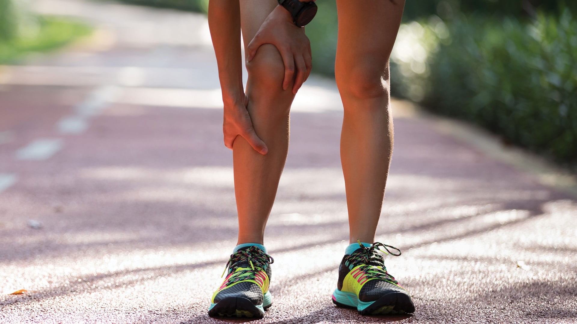
Calf Strain
A Calf Strain is tearing of the fibers of the muscle’s gastrocnemius or soleus. Most commonly they affect the middle portion of the gastrocnemius. Strains of the soleus muscle are less common. These can occur in adults who participate in activities that require quick acceleration and deceleration, as well as rapid changes in direction. This includes sports such as basketball and tennis.
Symptoms include pain in the back of the lower leg or behind the knee, with difficulty walking. There may be a tearing or popping sensation.
Treatment often includes a period of rest, activity modification, and physical therapy. Dr. Sampson uses ultrasound for accurate diagnosis supervises a return to activity.
Knee Arthritis
Knee arthritis occurs at the joint between the femur and tibia. Over time the cartilage wears down and causes the bones to rub together causing pain. There are many causes including wear and tear over time, trauma, infection, chronic injury, etc.
Symptoms include pain, weakness, and loss of motion. Patients may note catching, locking, or popping.
Treatment often includes a period of rest, activity modification, and physical therapy. Dr. Sampson uses ultrasound for guided injections in refractory cases.
Lateral Collateral Ligament (LCL) Injury
The Lateral Collateral Ligament (LCL) is one of the four ligaments that stabilize the knee. The LCL is located on the outer side of the knee, and it prevents the knee from collapsing outward. LCL sprains are may occur in sports that require quick side-to-side movements, such as hockey, skiing, soccer, and football. A sprain of the LCL can range from a mild injury to a complete tear of the ligament. Injury may occur when with contact on the inside of the knee or from a twisting motion on a bent knee.
Symptoms may include hearing or feeling a popping or tearing sensation along the outer side of knee. Pain along the outer part of the knee along with swelling and bruising. Painful movement or weight bearing. A feeling giving way in the knee, especially when attempting to quickly change direction
Treatment often includes a period of rest, bracing, activity modification, and physical therapy. Dr. Sampson uses ultrasound for accurate diagnosis and supervises a return to activity.
Medial Collateral Ligament (MCL) Injury
The Medial Collateral Ligament (MCL) is one of the four ligaments that stabilize the knee. The MCL is located on the inner side of the knee, and it prevents the knee from collapsing inward. MCL sprains are very common, especially in sports that require quick side-to-side movements, such as hockey, skiing, soccer, and football. A sprain of the MCL can range from a mild injury to a complete tear of the ligament. Injury may occur when with contact on the outside of the knee or from a twisting motion on a bent knee.
Symptoms may include hearing or feeling a popping or tearing sensation along the inner side of knee. Pain along the inner part of the knee along with swelling and bruising. Painful movement or weight bearing. A feeling giving way in the knee, especially when attempting to quickly change direction.
Treatment often includes a period of rest, bracing, activity modification, and physical therapy. Dr. Sampson uses ultrasound for accurate diagnosis and supervises a return to activity.
Meniscus Injury
The meniscus is a cartilage in the knee that acts as a shock absorber. There are two menisci in the knee, one on the inner aspect of the knee and one on the outer aspect of the knee. Meniscus tears can happen with a twisting knee injury, especially in younger athletes. Additionally degenerative tears, which can happen over time. Degenerative tears can start to occur as early as in the mid-thirties and might occur without any known injury. Meniscus tears can happen by themselves or with other injuries of the knee such as ligament tears.
Symptoms include pain and swelling of the knee. There may be locking or catching of the knee with movement.
Treatment often includes a period of rest, activity modification, and physical therapy. Dr. Sampson uses ultrasound for accurate diagnosis and guided injections in refractory cases.
Osgood-Schlatter Disease
Osgood-Schlatter Disease refers to a condition occurring during adolescence that causes pain, swelling and soreness on an area of the upper shinbone, just below the knee. The condition commonly occurs during a growth spurt. During this time the tibial tuberosity is vulnerable to overuse in an active teenager who is involved in a lot of running and jumping activities. The patellar tendon attaches to the tibial tuberosity and with repetitive activity can cause traction of this growth center causing inflammation. This can also be caused by repetitive activities in growing teenagers who do not allow enough time in between activities to heal.
Symptoms include pain at the bump below the knee with activity or after a fall. There may be swelling and enlargement of this bump on the upper shinbone. Forceful contraction of the thigh muscle can also cause pain. One or both knees may be affected.
Treatment often includes a period of rest, activity modification, and physical therapy. Dr. Sampson will confirm the diagnosis and supervise a safe return to activity.
Patellar Tendinopathy (Jumper’s Knee)
The patellar tendon attaches the kneecap to the shin bone. The patellar tendon works with the muscles at the front of the thigh to extend the knee. This injury is most common in athletes who frequently jump, such as when playing basketball and volleyball. However, even people who don’t participate in jumping sports can get patellar tendonitis.
Symptoms include knee pain, swelling, and stiffness. Pain is often present over the patellar tendon with direct palpation.
Treatment often includes a period of rest, activity modification, and physical therapy. Dr. Sampson uses ultrasound for accurate diagnosis and guided injections in refractory cases.
Patellofemoral Pain Syndrome (Runner’s Knee)
Patellofemoral Pain Syndrome is a common cause of knee pain. The kneecap sits in a groove at the bottom of the thigh bone. There are muscles, tendons, and ligaments surrounding the patella that control how the kneecap moves. If the structures surrounding the kneecap are out of proper balance, pain can develop based on the interaction of the kneecap and the thigh bone. Causes may include repetitive running and jumping activities, increase in training, fall on the front of the knee, muscle weakness, or malalignment of the upper or lower leg.
Symptoms include a dull, aching pain in the front of the knee. This pain may become worse when walking up or down stairs, kneeling or squatting, and sitting for long periods of time with the knee bent.
Treatment often includes a period of rest, activity modification, and physical therapy. Dr. Sampson uses ultrasound for accurate diagnosis and supervises a safe return to activity.
PCL Injury
A PCL injury occurs when there is damage to the posterior cruciate ligament, a major knee located inside the knee. The PCL is the main stabilizing ligament of the knee for front to back forces. In particular, the PCL prevents the lower leg from slipping too far back in relation to the upper leg, especially when the knee is flexed. Injuries may range from disruption of a few fibers to a complete tear of the ligament. The PCL most often is injured when the front of the knee hits the dashboard during an automobile accident. During sports activities, the PCL also can tear when an athlete falls forward and lands hard on a bent knee, which is common in football, basketball, soccer and especially rugby.
Symptoms may mild knee swelling, with or without the knee giving out with walking or standing, and with or without limitation of motion. This symptom may begin one to two weeks after the injury or even later.
Treatment often includes a period of rest and bracing followed by physical therapy. Dr. Sampson will confirm the diagnosis and supervises a safe return to activity.
Quadriceps Tendon Injury
The quadriceps muscle group is made up of four muscles in the front of the thigh. The quadriceps tendon attaches the quadriceps muscles to the kneecap. The patellar tendon attaches the kneecap to the shin bone. These muscles, tendons and bones work together to straighten the knee. Although patellar and quadriceps tendon ruptures or tears are rare, they are serious injuries. A rupture may occur in the middle of the tendon or close to the bone. Ruptures usually occur when the quadriceps muscle contracts forcefully while the knee is bent. The patellar tendon is more likely to rupture in athletes under age 40, while patients over age 40 are more likely to rupture the quadriceps tendon. Steroid use and certain diseases such as rheumatoid arthritis, lupus, diabetes and kidney failure increase the risk for patellar and quadriceps tendon ruptures. Tendinopathy, previous knee surgeries and degenerative changes also increase the risk for rupture.
Symptoms may include hearing or feeling a pop, and the athlete may not be able to actively extend the knee. The knee may feel unstable and is often swollen and painful.
Treatment often includes surgical intervention for full tears. Dr. Sampson uses ultrasound for accurate diagnosis to expedite treatment. For partial tears or tendinopathy, treatment often includes a period of rest, activity modification, and physical therapy.
Sinding-Larsen Johansson Disease
Sinding-Larsen Johansson Disease is a cause of knee pain in children. Most commonly seen between the ages of 8-to-13-year-old active individuals. Children often complain of pain and swelling at the bottom of the kneecap that may be worsened by climbing stairs, running, jumping, deep bending of the knee, or kneeling. Children may develop pain during periods of increased activities or periods of rapid growth. The cause is a painful irritation of the growth plate at the bottom of the kneecap. Children have open growth areas on their bones that are made of cartilage where muscles and tendons attach. These growth areas can become irritated with repetitive stress or a sudden blow to the area.
Symptoms include pain and swelling at the bottom of the kneecap that may be worsened by climbing stairs, running, jumping, deep bending of the knee, or kneeling. They may have swelling or a bump at the bottom of the kneecap.
Treatment often includes a period of rest, activity modification, and physical therapy. Dr. Sampson will confirm the diagnosis and supervise a safe return to activity.
Schedule your evaluation with a Board Certified Sports Medicine Physician today!
I will come up with a diagnosis and treatment plan during your first appointment.
This website uses cookies to improve your experience. We'll assume you're ok with this, but you can opt-out if you wish. Cookie settingsACCEPT
Privacy & Cookies Policy
Privacy Overview
This website uses cookies to improve your experience while you navigate through the website. Out of these cookies, the cookies that are categorized as necessary are stored on your browser as they are as essential for the working of basic functionalities of the website. We also use third-party cookies that help us analyze and understand how you use this website. These cookies will be stored in your browser only with your consent. You also have the option to opt-out of these cookies. But opting out of some of these cookies may have an effect on your browsing experience.
Necessary cookies are absolutely essential for the website to function properly. This category only includes cookies that ensures basic functionalities and security features of the website. These cookies do not store any personal information.